You are not your MRI
What is an MRI?
It’s a common misconception within the general non-medical population that an MRI scan paints a 1000 words. This isn’t always the case. Before we get to that, lets talk about what an MRI is. It stands for Magnetic Resonance Imaging. It cleverly uses magnetic fields and radio waves to produce detailed pictures of bones, soft tissues and organs without the need for surgery. This is the form of imaging that is considered the crème de la crème. They show much more detail, including many structures that are not visible on an X-ray. Another bonus is that the person having an MRI isn’t exposed to the radiation that occurs with X-rays or CT scans.
Now I’ve covered some pros, and there are certainly ones when used in the correct situations, let’s go into the nitty gritty of why they aren’t quite as good as you may think for musculoskeletal problems.
Will my pain be found on an MRI?
MRIs are not pain scans! I’m going to say that again… MRI are not pain scans! I say this a lot to my patients in clinic. When we send someone for an MRI of a certain body part, the images don’t come back with a big arrow that points to where the pain is unfortunately. I really wish it was as simple as that.
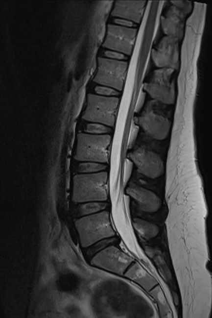
The MRI is just a still life photograph showing structural changes to the area. Some of those findings you are better off not knowing. They can be completely incidental and not linked to your symptoms. Our bodies move and work in a very functional way. The MRI misses this in it’s still life assessment. It is actually taken whilst you are lying down. Missing the weight bearing element that gravity will create. Interestingly there are some new MRI facilities that have now started doing standing MRIs. This would be better in assessing most spinal complaints with the disc compression changing when upright.
Don’t get me wrong MRIs are an excellent tool for ruling out nasty things. But in terms of locating pain… it definitely shouldn’t be the first line of defence when assessing. An MRI should only be ordered when we suspect something more sinister is going on, no progress is being made with conservative care or the treatment plan is about to change by referring for surgical intervention. Research has shown that once you have an MRI, you are far more likely to end up having surgery. This is a complicated matter because there will obviously be a few factors involved with this connection. The one that is continuously formed from the researchers is that it’s because the image and physical findings are imprinted into your mind and psychologically creates a permanent picture of how you think of your body. For example, you’ll curse yourself with having a “bad back” forever more, just because you saw some wear in your spine, which might not have been the cause of your pain in the first place.
How does a physical examination help?
A physical examination is a much more useful tool in determining where the source of the pain is actually coming from. There is a huge range of orthopaedic and neurological testing that can be done. I always warn my patients during a consultation that the testing is meant to be pain provoking. This allowing me to know exactly where the pain is coming from. Creating a much better picture of what’s going on. So if you do have an MRI it should be used along with your presentation and an examination to determine what the symptomatic problem is.
Let me give you an example of where an examination can be so important. A pain that starts in the groin and refers down the front of the thigh could be coming from a number of places. This includes the spine, pelvis joint, hip or knee. Crazy right? Now lets say an MRI is warranted in this case . I’m suspicious of something more sinister. How would I know what area to image without a physical examination to find out where the source of the pain is. That could be a huge waste of resources if the wrong area was imaged. AND it shows an incidental finding that has nothing to do with the current pain. This could lead to unnecessary surgery, which won’t improve the pain because guess what? It’s the wrong joint! Don’t be so shocked… it happens!
An interesting fact is that people can have structural changes present on an MRI, but absolutely no pain! So that leads on to the question… What exactly is pain? That’s a whole other conversation. It is still somewhat of a mystery to the medical profession. Pain is complex, multi-factorial and is why the answer is not usually found on an MRI scan. What I’m saying is that you could have an identical looking MRI scan to someone who is the same age and had a similar life, showing certain bits of wear and tear to the muscles, joints and bones, BUT you have pain and the other person doesn’t! This is where other factors can be involved that over sensitise the nerve endings. These factors can come in the form of inflammation, stress, hormones, and neurotransmitters. This can’t be seen on an MRI.
Let’s talk research…
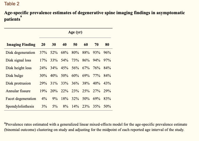
A huge study was done on numerous people that did NOT have pain. They were given an MRI on their spine to see the features that were present. The results are amazing and very reassuring that you should be able to return to a pain free life, even if you have a symptomatic version of these findings!
A number of findings are presented in a table above, but lets talk about the “problem” that most of you will be familiar with, a disc bulge. The research showed that the chances of you having a disc bulge in your 30s is 30%. Remember this is in individuals who do not have any pain! As we get older, the rate of this finding increases dramatically. When you are in your 70s, the chances increase to 77%! 77% of people WITHOUT back pain have disc bulges when they are 70. If that statement doesn’t normalise the presence of disc bulges, I don’t know what does. Obviously the pain that can come with them is a different matter, but if you’ve been diagnosed with a disc bulge, I really don’t want you to think you are doomed to a lifetime of pain!
Now we are at the end of the article, it may appear that I do not believe in MRI scans. This certainly isn’t the case. In fact, I send on average about 3 patients per month for an MRI scan to gain a deeper understand of what is going on underneath the skin. This is always done after a full consultation, collecting a history of the problem and a thorough physical examination to locate the source of the pain. We use The Cobalt Clinic which is an excellent facility run as a charity that we are lucky enough to have based in Cheltenham. This allows self funding patients to only pay half of what an MRI would normally cost, as they contribute towards it. At the time of this being written, it costs £266 for one body part to be imaged and reported on. You will never be left alone with the result, as your referrer will talk you through the findings, linking it to your presentation and examination to give the most likely cause of your symptoms.
Read onto our next article by Chiropractor Kelly on Acupuncture.


